Gallery of images
Art and Science : Gallery of images
Neurodevelopment meets Statistical Analysis, Deep Learning classification and algorithms
Microglia cell classification in human embryo : generated by T. Perochon and D.Holcman
— quote our reference for private or public usage "T. Perochon, Z. Krsnik, T. Lagache, D.A. Menassa D. Holcman, Spatiotemporal mapping of human microglia during brain development with advanced spatial statistics assisted by deep-learning, pre-print."
Pictures
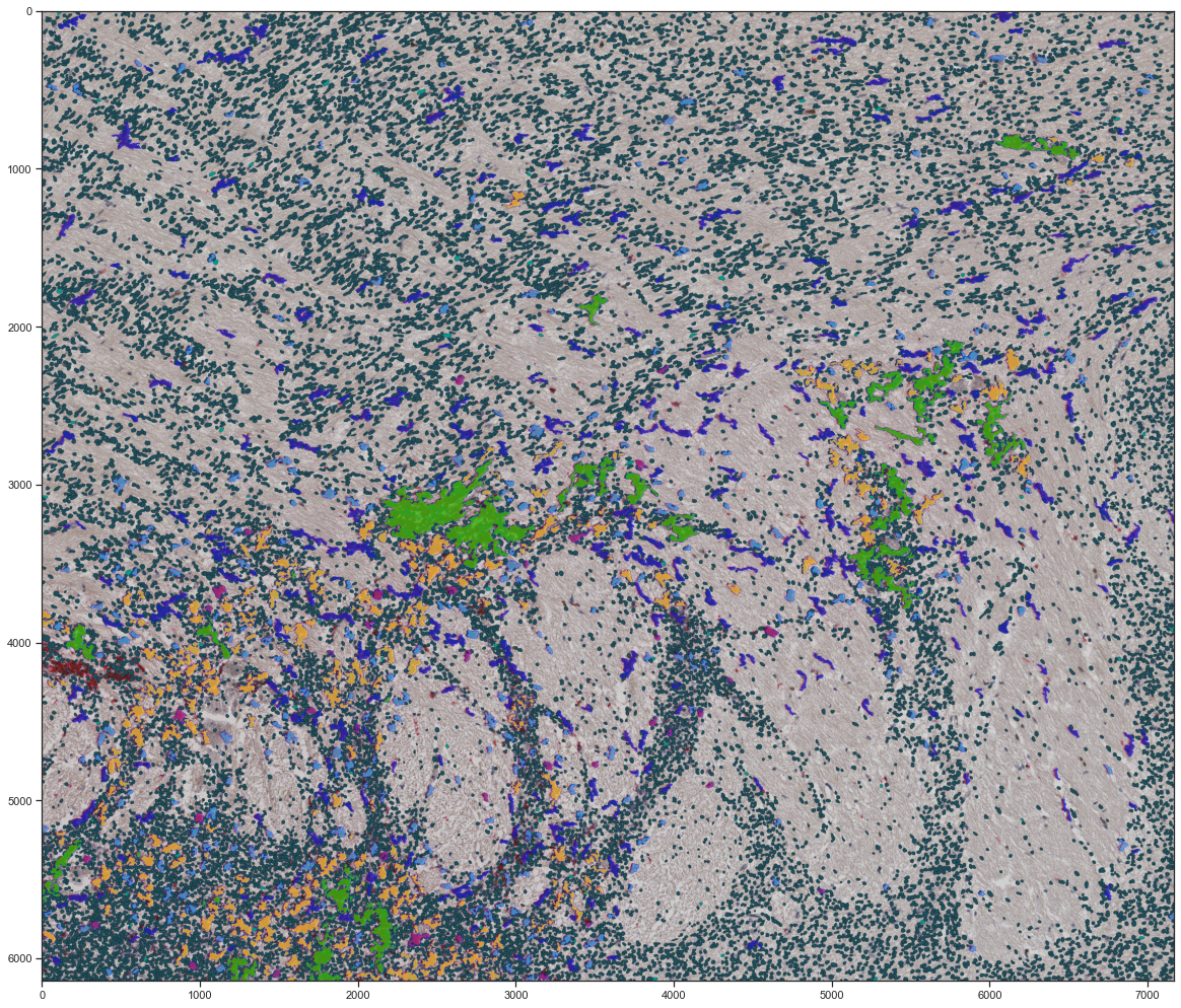
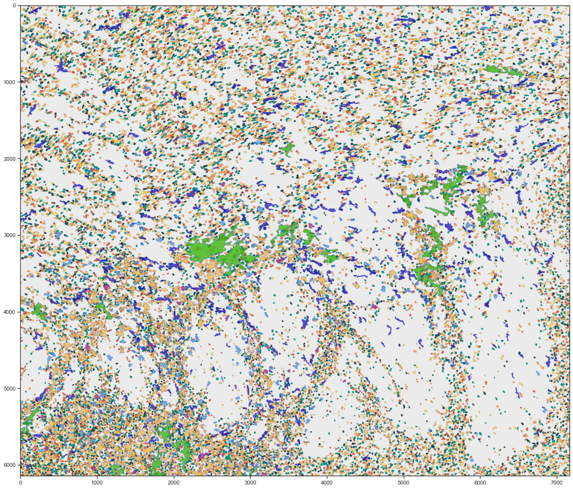
Legend fig.1 :Cell distributed in the Striatum, colored by cell types using deep learning classification.
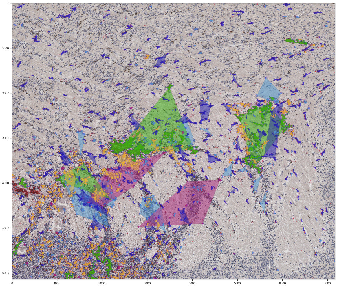
Legend fig.2 :Variant of the previous image with convex hulls are obtained by an optimized version of the (DBSCAN) clustering algorithm.
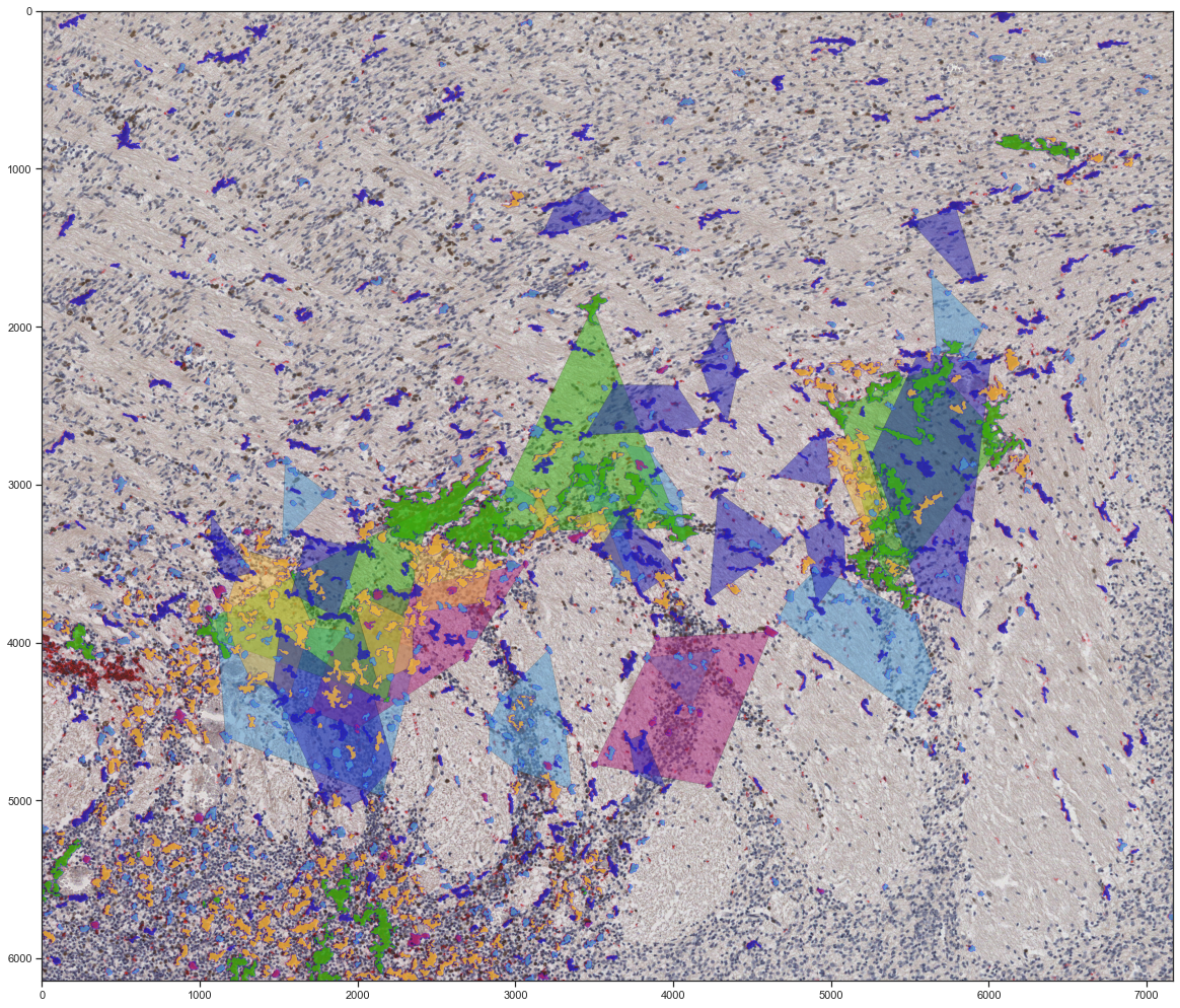
Legend fig.3 :Variant of the previous image

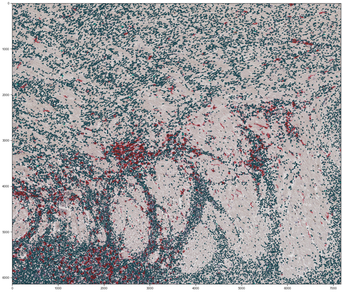
Legend fig.4 : Cellpose analysis
Legend fig.5 : Cell distributed in the Striatum, colored by cell types and convex hull segmentations
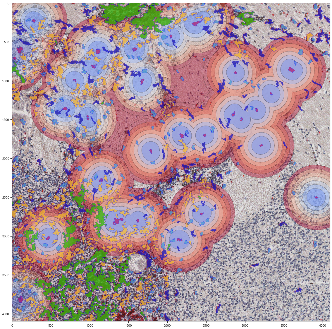
Legend fig.6 :Spatial statistics based on levelset analysis
Legend fig.7 :Microglia automated segmentation using deeplearning with cellpose classification
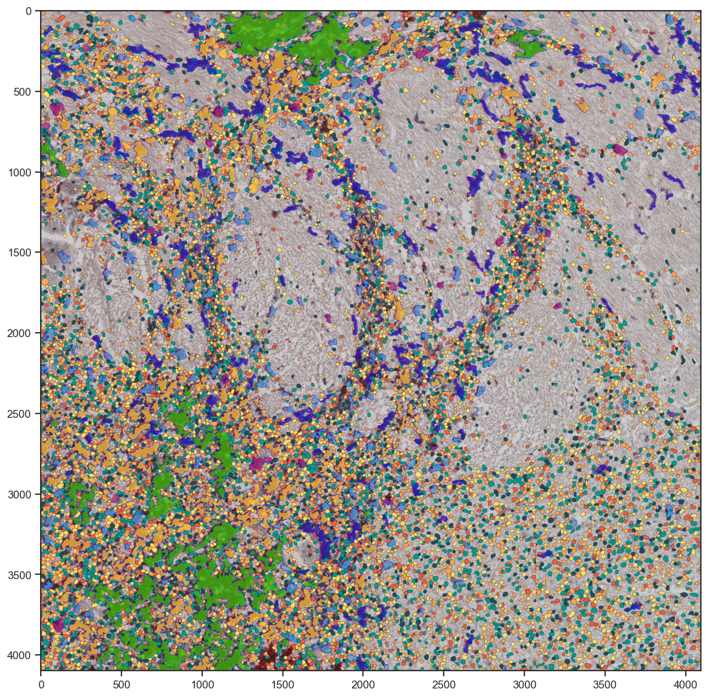
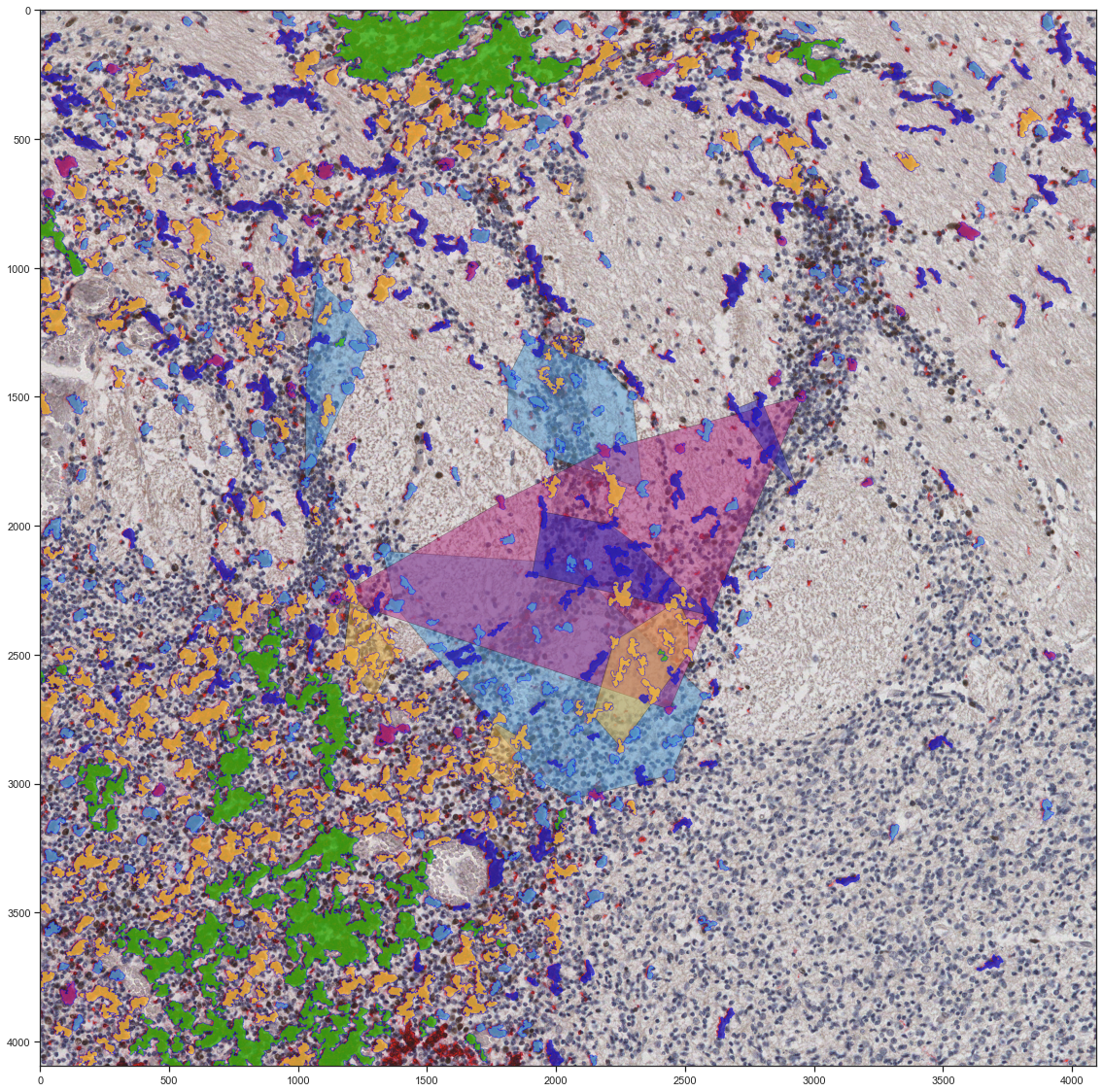
Legend fig.8 :Magnification of Striatum with cellpose classification and deep learning classification
Legend fig.9 :Cell pose superimposed with our algorithm for microglia present in the Striatum